

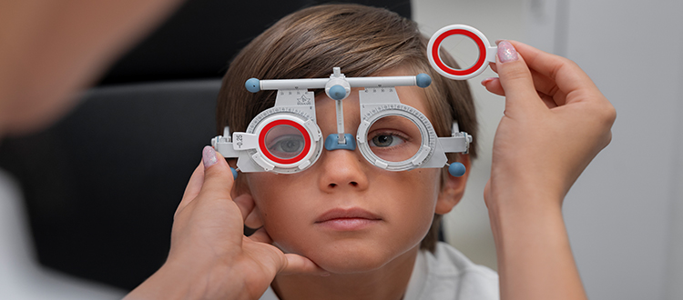
05 Nov Where to Get an Optic Nerve OCT and Visual Field Test Done in India
Last updated: 4 November 2026 | Author: Prashant Tyagi (Stem Cell Biotechnology Specialist, 10+ years) and Dr. Pallavee Senior Consultant | Ophthalmologist & Eye Surgeon 22+ Years Experience
If you or your child has been told the optic nerve is damaged, two tests do most of the work: OCT of the optic nerve and visual field testing. Together they show how much nerve fibre is left and how much of your vision it supports. This guide explains what the tests measure, where to get them in India, and how to make sure the results are useful.
At a glance
- OCT measures the thickness of the nerve fibre layer, and visual field testing maps the area you can see
- The tests are painless and done at eye hospitals and clinics, usually with an ophthalmologist’s request
- Ask for the full printout and the digital files, not only a one-line report
- Repeat tests on the same type of machine, so changes can be compared fairly
- These tests help find the cause and track change, and they do not by themselves restore vision
Quick Navigation
- What the tests measure
- Where to get them in India
- Questions to ask before you book
- How to prepare
- Getting reliable results
- Reading your report
- What comes next
- Frequently asked questions
What the Tests Measure
| Test | What it shows | Notes |
|---|---|---|
| OCT of the optic nerve | Thickness of the retinal nerve fibre layer around the optic nerve, and sometimes the ganglion cell layer at the macula | Quick, painless, no contact with the eye |
| Visual field test (perimetry) | Which parts of your vision are reduced or missing | Needs concentration; usually several minutes per eye |
| Colour vision test | How well colours are recognised | Often reduced in optic nerve disease |
| Visual evoked potentials | How quickly signals travel from the eye to the brain | Done in selected cases |
The tests answer different questions. OCT shows structure, meaning how much nerve is left, while the visual field shows function, meaning what you can actually see. Neither explains the cause by itself, which is why an MRI and other tests may also be needed. See our pages on optic atrophy and optic neuritis.
Where to Get Them in India
- Tertiary eye hospitals and eye institutes often offer OCT, perimetry and neuro-ophthalmology under one roof. Examples include AIIMS in Delhi, L V Prasad Eye Institute in Hyderabad, Sankara Nethralaya in Chennai and the Aravind Eye Hospitals. This is not a ranking or endorsement, and services vary.
- Medical college hospitals and government eye departments may provide these tests at lower cost, sometimes with a waiting list.
- Private eye clinics and retina centres commonly have OCT, and many have automated perimetry.
- Diagnostic centres may offer imaging, but make sure an ophthalmologist, ideally with neuro-ophthalmology experience, reviews the results.
You can also see our list of optic nerve specialist doctors.
Questions to Ask Before You Book
- Do you perform OCT of the optic nerve (retinal nerve fibre layer) and automated visual field testing?
- Do I need a referral or an appointment, and how long will the visit take?
- Do I need dilating drops, and should someone come with me?
- Will I receive the full printouts and digital files?
- Can I repeat the tests on the same machine for follow-up?
- Is the test reviewed by a doctor with optic nerve experience?
How to Prepare
- Bring your glasses, earlier reports, MRI results and a list of medicines
- If dilating drops are used, expect blurred near vision for a few hours, so bring a companion
- For the visual field test, rest well, eat normally and wear the correct glasses if advised
- Tell the technician if you feel tired, since it affects the result, and ask for a break if needed
- For children, ask the centre whether they use child-friendly methods
Getting Reliable Results
Visual field tests can vary with attention, tiredness and learning effects, so one test is rarely enough. Many specialists repeat the test to confirm a finding. For OCT, good image quality matters: a poorly centred or blurred scan can give misleading numbers. Ask whether the report shows quality indicators, and request a repeat if the scan looks unreliable. Do follow-up tests on the same type of machine whenever possible, because numbers from different devices are not always directly comparable.
Reading Your Report
- OCT: The report usually compares your nerve fibre thickness with an age-matched reference range, often in colour-coded sectors. Thinner values suggest fibre loss, but they must be read with your eye examination and history.
- Visual field: The report shows maps of reduced sensitivity and summary numbers. Reliability indicators tell the doctor how trustworthy the test was.
- Keep every printout, label the eye (right or left) and date, and bring them to every visit.
What Comes Next
If the tests show optic nerve damage, the next step is to find the cause, such as compression, inflammation, glaucoma, nutritional deficiency, a hereditary condition or injury, because treating the cause early can protect remaining vision. Fibres that have died do not regrow, and stem cell and nerve regeneration approaches remain investigational. Read our page on optic atrophy treatment in India, and our guide to living with partial vision loss.
Frequently Asked Questions
Is an OCT painful?
No. It is a quick, non-contact scan.
How long does a visual field test take?
Usually several minutes per eye, plus preparation time.
Do I need to dilate my eyes?
Sometimes. Your clinic will tell you in advance.
How often should the tests be repeated?
Your specialist will decide. At first, tests may be repeated every few months, then less often if stable.
Can a child have these tests?
Many children can, depending on age and cooperation. Centres use age-appropriate methods.
Do the tests show the cause of optic atrophy?
Not alone. They show how much damage there is, and other tests such as MRI and blood tests help find the cause.
Can I get a second opinion on my reports?
Yes. Share your reports and images for an expert review before making treatment decisions.
More Official Sources
- National Eye Institute (NEI), NIH
- National Institute of Neurological Disorders and Stroke (NINDS), NIH
- NIH Genetic and Rare Diseases Information Center (GARD)
- World Health Organization – Blindness and Vision Impairment
- Indian Council of Medical Research (ICMR)
- Ministry of Health and Family Welfare (MoHFW)
Related Reading
- Optic Atrophy Treatment in India
- Optic Neuritis
- Leber Hereditary Optic Neuropathy
- Glaucoma Optic Atrophy
This article is for general information and does not replace an examination by a qualified eye specialist.

Sorry, the comment form is closed at this time.